2.7 Cell - the genetic unit of the living. Chromosomes, their structure (shape and size) and functions
Video lesson 1: Cell division. Mitosis
Video lesson 2: Meiosis. Phases of meiosis
Lecture: A cell is the genetic unit of a living thing. Chromosomes, their structure (shape and size) and functions
The cell is the genetic unit of life
The single cell is recognized as the basic unit of life. It is at the cellular level that processes occur that distinguish living matter from non-living matter. Each of the cells stores and intensively uses hereditary information about the chemical structure of proteins that must be synthesized in it, and therefore it is called the genetic unit of the living. Even nuclear-free erythrocytes at the initial stages of their existence have mitochondria and a nucleus. Only in their mature state do they lack the structures for protein synthesis.
To date, science does not know cells that would not contain DNA or RNA as a carrier of genomic information. In the absence of genetic material, the cell is not capable of protein synthesis, and therefore life.
DNA is present not only in the nuclei, its molecules are contained in chloroplasts and mitochondria, these organelles can multiply inside the cell.
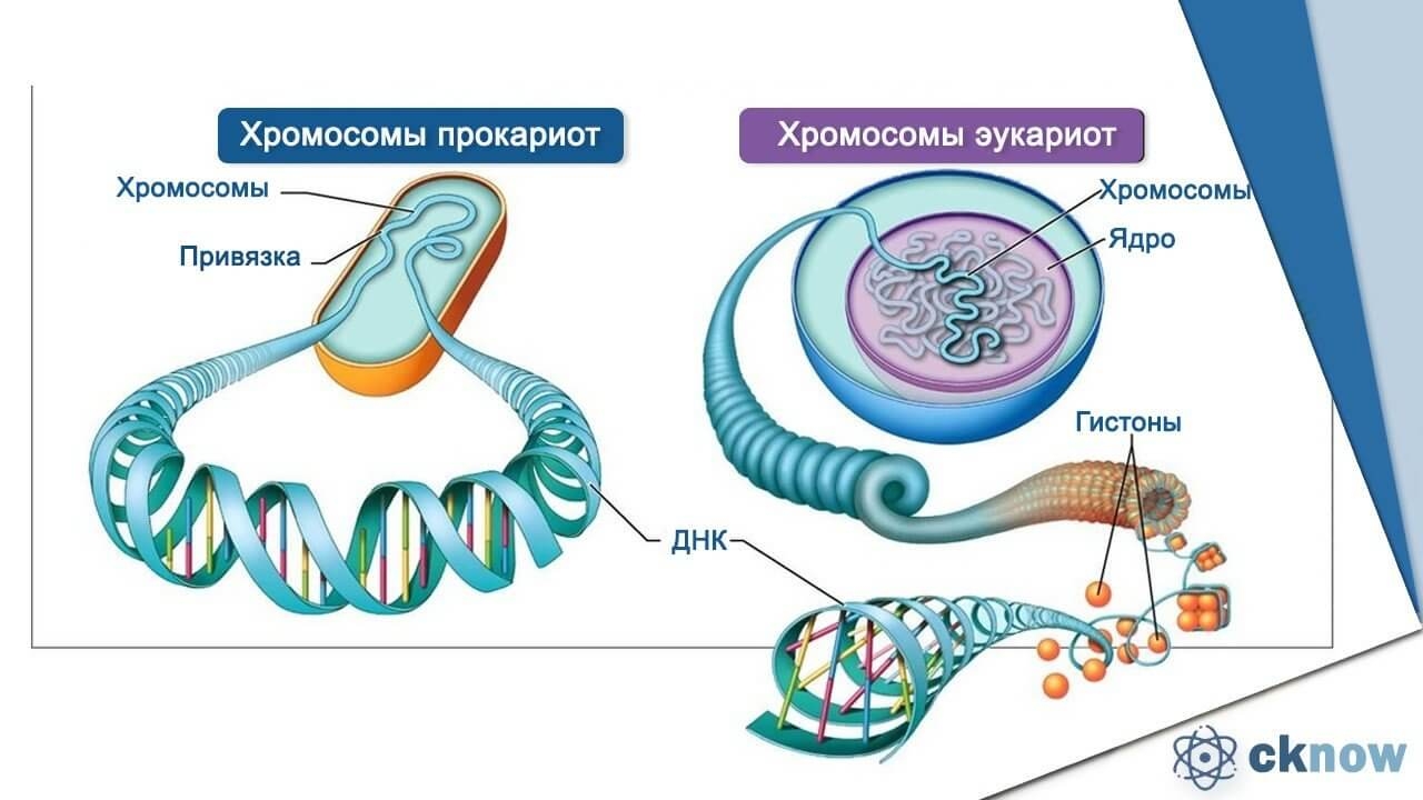
DNA in a cell is in the form of chromosomes - complex protein-nucleic acid complexes. Eukaryotic chromosomes are located in the nucleus. Each of them is a complex structure of:
The only long DNA molecule, 2 meters of which is packed into a compact structure (in humans) up to 8 microns in size;
Special histone proteins, whose role is to pack chromatin (the substance of the chromosome) into a familiar rod-shaped form;
Chromosomes, their structure (shape and size) and functions
This dense packing of genetic material is produced by the cell before division. It is at this moment that densely packed, formed chromosomes can be examined under a microscope. When DNA is folded into compact chromosomes called heterochromatin, messenger RNA synthesis is not possible. During the period of cell mass recruitment and its interphase development, chromosomes are in a less packed state, which is called interchromatin and mRNA is synthesized in it, DNA replication occurs.
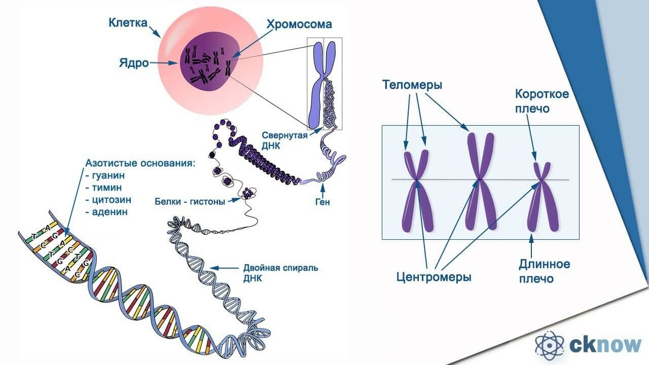
The main elements of the structure of chromosomes are:
centromere. This is a part of the chromosome with a special sequence of nucleotides. It connects two chromatids together, participates in conjugation. It is to it that the protein filaments of the spindle tubes of cell division are attached.
Telomeres. These are the terminal sections of chromosomes that are not capable of connecting with other chromosomes, they play a protective role. They consist of repeating sections of specialized DNA that form complexes with proteins.
Points of initiation of DNA replication.
Chromosomes of prokaryotes are very different from eukaryotic ones, representing DNA-containing structures located in the cytoplasm. Geometrically, they represent a ring molecule.
The chromosome set of a cell has its own name - a karyotype. Each of the species of living organisms has its own composition, number and shape of chromosomes, characteristic only for it.
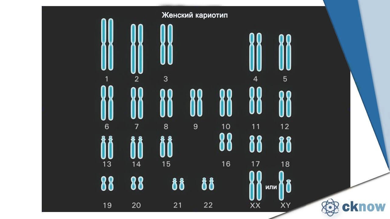
Somatic cells contain a diploid (double) chromosome set, half of each parent received.
Chromosomes responsible for encoding the same functional proteins are called homologous. The ploidy of cells can be different - as a rule, in animals, gametes are haploid. In plants, polyploidy is now a fairly common phenomenon, which is used to create new varieties as a result of hybridization. Disruption of the amount of ploidy in warm-blooded animals and humans causes serious congenital diseases such as Down's syndrome (the presence of three copies of the 21st chromosome). Most often, chromosomal abnormalities lead to the non-viability of the organism.
In humans, the complete chromosome set consists of 23 pairs. The largest known number of chromosomes, 1600, was found in the simplest planktonic organisms, radiolarians. The smallest set of chromosomes in the Australian black bulldog ants is only 1.
The life cycle of a cell. Phases of mitosis and meiosis
Interphase, in other words, the length of time between two divisions is defined by science as the life cycle of a cell.
During the interphase, vital chemical processes take place in the cell, it grows, develops, and accumulates reserve substances. Preparation for reproduction involves doubling the content - organelles, vacuoles with nutritional content, the volume of the cytoplasm. It is thanks to division, as a way to rapidly increase the number of cells, that long life, reproduction, an increase in the size of the body, its survival in case of injuries and tissue regeneration are possible. The following stages are distinguished in the cell cycle:
Interphase. Time between divisions. First, the cell grows, then the number of organelles increases, the volume of the reserve substance increases, proteins are synthesized. In the last part of the interphase, the chromosomes are ready for the subsequent division - they consist of a pair of sister chromatids.
Mitosis. This is the name of one of the methods of nuclear division, characteristic of bodily (somatic) cells, in its course, 2 cells are obtained from one, with an identical set of genetic material.
Meiosis is characteristic of gametogenesis. Prokaryotic cells have retained the ancient method of reproduction - direct division.
Mitosis consists of 5 main phases:
- Anaphase. The efforts of the microtubules break off the connections of the chromosomes in the centromere region, and with force stretch them to the poles of the cell. In this case, the chromosomes sometimes take a V-shape due to the resistance of the cytoplasm. A ring of protein fibers appears in the region of the metaphase plate.
- Telophase. Its beginning is considered the moment when the chromosomes reach the poles of division. The process of restoring the internal membrane structures of the cell begins - EPS, the Golgi apparatus, the nucleus. Chromosomes unpack. The nucleoli assemble and the synthesis of ribosomes begins. The spindle of division disintegrates.
- cytokinesis. The last phase, in which the protein ring that appeared in the central region of the cell begins to shrink, pushing the cytoplasm towards the poles. There is a division of the cell into two and the formation of the protein ring of the cell membrane in place.
Prophase. Its beginning is considered the moment when the chromosomes become so densely packed that they are visible under a microscope. Also, at this time, the nucleoli are destroyed, a division spindle is formed. Microtubules are activated, the duration of their existence decreases to 15 seconds, but the rate of formation also increases significantly. Centrioles diverge to opposite sides of the cell, forming a huge number of constantly synthesizing and decaying protein microtubules that extend from them to the centromeres of chromosomes. This is how the spindle is formed. Membrane structures such as ER and the Golgi apparatus disintegrate into separate vesicles and tubules randomly located in the cytoplasm. Ribosomes are separated from the ER membranes.
metaphase. A metaphase plate is formed, consisting of chromosomes balanced in the middle of the cell by the efforts of opposite centriole microtubules, each pulling them in its own direction. At the same time, the synthesis and disintegration of microtubules continues, their kind of "bulkhead". This phase is the longest.
Regulators of the process of mitosis are specific protein complexes. The result of mitotic division is a pair of cells with identical genetic information. In heterotrophic cells, mitosis proceeds faster than in plant cells. In heterotrophs, this process can take from 30 minutes, in plants - 2-3 hours.
To generate cells with half the normal number of chromosomes, a different division mechanism is used by the body - meiosis.
It is associated with the need to produce germ cells; in multicellular organisms, it avoids a constant doubling of the number of chromosomes in the next generation and makes it possible to obtain new combinations of allelic genes. It differs in the number of phases, being longer. The resulting decrease in the number of chromosomes leads to the formation of 4 haploid cells. Meiosis is two divisions that follow each other without interruption.
The following phases of meiosis are defined:
Prophase I. Homologous chromosomes approach each other and unite longitudinally. Such an association is called a conjugation. Then there is a crossing over - double chromosomes cross their shoulders and exchange sections.
Metaphase I Chromosomes separate and take up positions at the equator of the cell spindle, taking on a V-shape due to microtubule tension.
Anaphase I Homologous chromosomes are stretched by microtubules to the poles of the cell. But unlike mitotic division, they diverge as whole, not as individual chromatids.
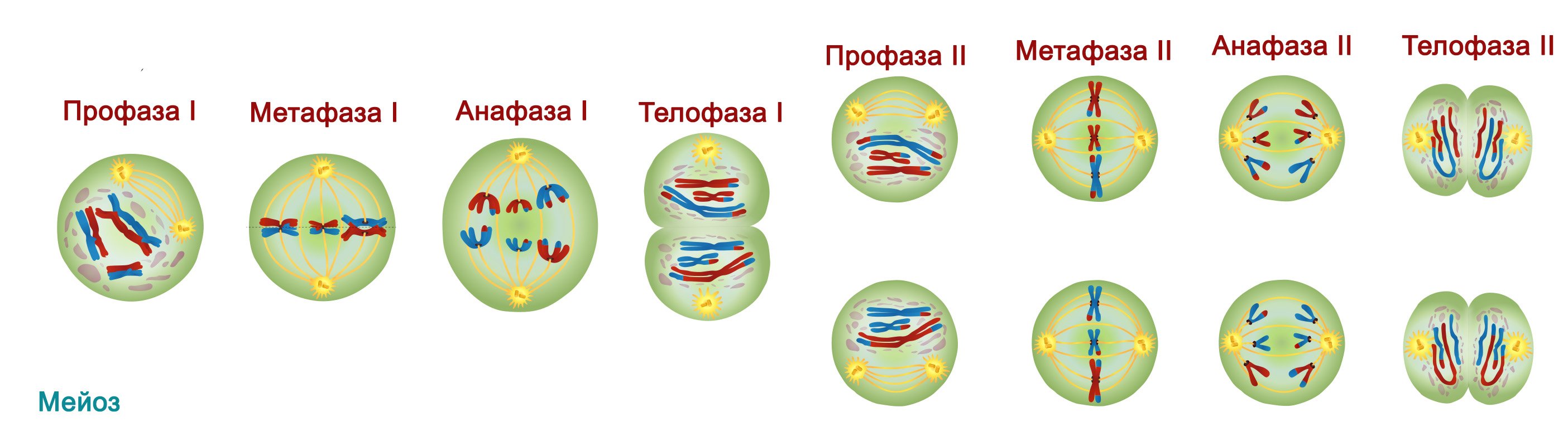
The result of the first division of meiosis is the formation of two cells with half the number of whole chromosomes. Between divisions of meiosis, interphase is practically absent, doubling of chromosomes does not happen, they are already two-chromatid.
Immediately following the first repeated meiotic division is completely similar to mitosis - in it, the chromosomes are divided into separate chromatids, distributed equally between new cells.
oogonia go through the stage of mitotic reproduction at the embryonic stage of development, so that the female body is already born with an unchanged number of them;
spermatogonia are capable of reproduction at any time during the reproductive period of the male body. Much more of them are generated than female gametes.
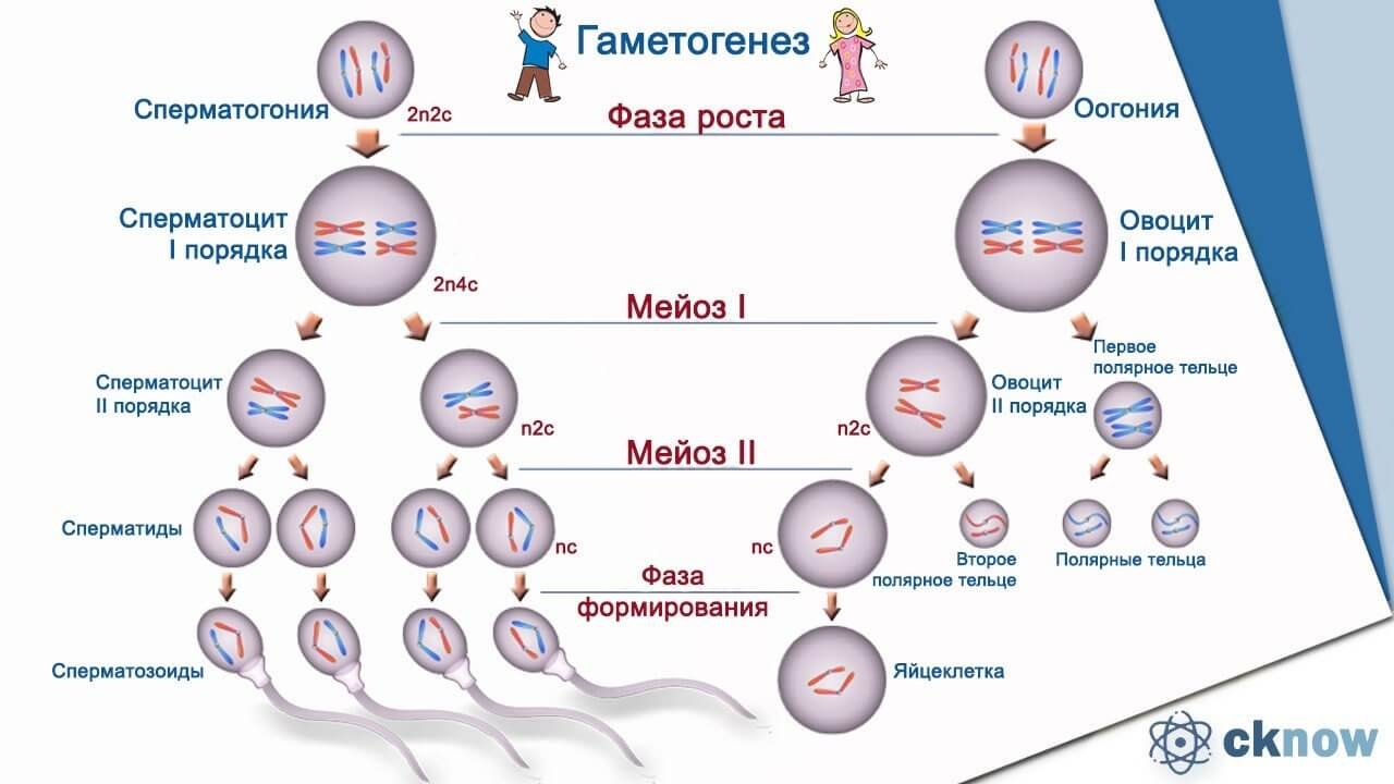
Gametogenesis of animal organisms occurs in the sex glands - gonads.
The process of transformation of spermatogonia into spermatozoa occurs in several stages:
Mitotic division transforms spermatogonia into spermatocytes of the 1st order.
As a result of a single meiosis, they turn into spermatocytes of the 2nd order.
The second meiotic division produces 4 haploid spermatids.
There is a period of formation. In the cell, the nucleus is compacted, the amount of cytoplasm decreases, and the flagellum is formed. Also, proteins are stored and the number of mitochondria increases.
The formation of eggs in an adult female body occurs as follows:
From the oocyte of the 1st order, of which there is a certain amount in the body, as a result of meiosis, with a decrease in the number of chromosomes by half, oocytes of the 2nd order are formed.
As a result of the second meiotic division, a mature egg and three small reduction bodies are formed.
This non-equilibrium distribution of nutrients between 4 cells is designed to provide a large resource of nutrients for a new living organism.
The eggs of ferns and mosses are produced in archegoniums. In more highly organized plants - in special ovules located in the ovary.
| | |